
In the section below, you will find representative pathology images of DICER1-related tumours.
Annotations provide defining characteristics of each tumor type.
- Ciliary Body Medulloepithelioma (CBME)
- Embryonal Rhabdomyosarcoma of the Uterine Cervix (cERMS)
- Paediatric Cystic Nephroma (pCN)
- Multi-nodular thyroid lesion, preference for follicular variant of papillary thyroid carcinoma (FVPTC)
- Nasal Chondromesenchymal Hamartoma (NCMH)
- Ovarian Embryonal Rhabdomyosarcoma (oERMS)
- Pineoblastoma (PinB)
Ciliary Body Medulloepithelioma (CBME)

Annotations: The tumor is adherent to the amorphous lens (below) and consists of palissadic glandular formations surrounded by hyaline stroma. Immature teratomatous stroma is also present as well as cartillagenous tissue (not shown), making this a teratomatous CBME.
Embryonal Rhabdomyosarcoma of the Uterine Cervix (cERMS)
 Annotations: Proliferation of small blue cells. A cambium layer is visible beneath the epithelium.
Paediatric Cystic Nephroma
 Annotations: Paediatric cystic nephroma showing septa without blastematous cells.

Annotations: Paediatric cystic nephroma showing septa without blastematous cells.
Multi-nodular Thyroid lesion, preference for follicular variant of papillary thyroid cancer (FVPTC)

Annotations: Follicles presenting characteristic papillary thyroid carcinoma nuclei: clear nuclei with grooves, pseudo-inclusion.
Nasal Chondromesenchymal Hamartoma (NCMH)

Annotations: Fibromyxoid tissue with abrupt cartilaginous nodules.
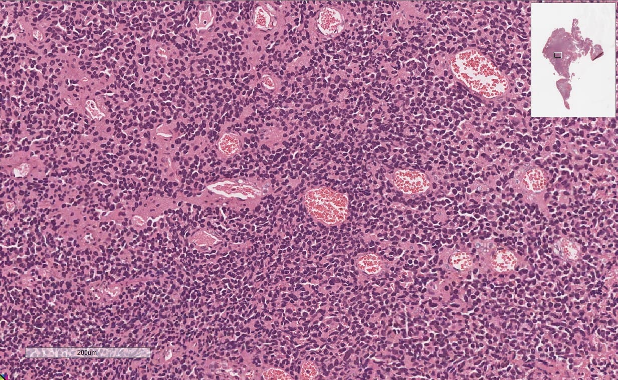
Ovarian Embryonal Rhabdomyosarcoma (oERMS)

Annotations: Proliferation of blue cells with scant cytoplasm. Presence of numerous mitoses.
Pineoblastoma (PinB)
Annotations: This embryonal tumor consists of monomorphous small blue cells without anaplasia, occasionally forming Homer-Wright rosettes.
This section was made possible thanks to the collaboration of Dr. Dorothee Dal Soglio and Dr. Benjamin Ellezam from the CHU Sainte-Justine.